Microscopes have revolutionized Dissecting Microscope how we see the microscopic world, playing a critical role in biology, forensics, botany, and other scientific fields. Among the many types of microscopes, the dissecting microscope, also known as a stereomicroscope, holds a unique place. Unlike compound microscopes designed for viewing thinly sliced specimens at high magnification, dissecting microscopes offer a three-dimensional view of larger, solid specimens at lower magnifications.
This article will explore the structure, working principle, applications, advantages, and limitations of dissecting-microscopes, helping you understand why this tool is invaluable in many scientific and educational settings.
What Is a Dissecting Microscope?
A dissecting microscope is an optical microscope specifically designed for low-magnification observation of a specimen. It provides a 3D view of the specimen using separate optical paths for each eye. This feature distinguishes it from standard microscopes and makes it ideal for observing surface details, manipulating small objects, and performing tasks like dissection and circuit board inspection.
Dissecting microscopes typically have a magnification range from 10x to 40x, though some models can go up to 100x. The name “dissecting” comes from its historical use in dissecting small organisms or anatomical parts under magnification.
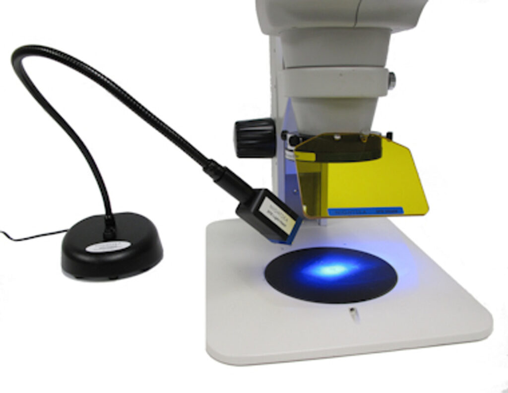
Components of a Dissecting Microscope
Dissecting microscopes consist of several key parts:
- Eyepieces (Ocular lenses): Usually two eyepieces providing stereoscopic (3D) vision.
- Objective lenses: These determine the magnification and are often mounted on a rotating turret.
- Zoom control knob: Allows the user to change magnification continuously within a set range.
- Focusing knob: Moves the head up and down to bring the specimen into sharp focus.
- Stage plate: The flat platform where the specimen is placed. It may be frosted or black/white to enhance contrast.
- Illuminators: Most dissecting microscopes have incident light (top lighting for surface features) and transmitted light (bottom lighting for semi-transparent objects).
- Stand or base: Provides stability and houses the light source in some models.
How a Dissecting Microscope Works
Unlike compound microscopes that use a single optical path, dissecting-microscopes have two separate optical channels. Each eye receives a slightly different image, which the brain combines to form a stereoscopic (3D) view. This perception of depth allows users to manipulate the specimen—such as dissecting, sorting, or assembling—while still under magnification.
The microscope’s lighting is equally essential. The incident (reflected) light highlights surface features, while transmitted light can be useful for slightly translucent specimens. The combination of these light sources can be adjusted depending on the needs of the user.
Applications of Dissecting Microscopes
Dissecting microscopes are used in a wide array of professional, academic, and industrial settings:
a. Biology and Botany
- Examining plant structures, insects, and animal specimens.
- Dissecting small organisms such as frogs, insects, or fish embryos.
b. Forensics
- Inspecting evidence such as hair, fibers, bullet casings, or other trace materials.
c. Electronics
- Soldering and inspecting circuit boards and miniature components.
d. Entomology and Taxonomy
- Observing and classifying insects and small organisms based on external features.
e. Education
- Teaching students about biological structures and dissection techniques.
f. Quality Control
- Inspecting manufactured goods, textiles, jewelry, and more for defects.
Advantages of a Dissecting Microscope
a. Three-Dimensional Imaging
The biggest advantage is the ability to view specimens in 3D, which is crucial for manipulation and dissection.
b. Ease of Use
Dissecting-microscopes require minimal sample preparation. Users can view specimens in their natural state without the need for slicing or staining.
c. Dual Lighting
The combination of incident and transmitted light offers versatility for examining different kinds of specimens.
d. Real-Time Manipulation
With ample working distance between the lens and the stage, users can manipulate specimens (e.g., dissect, sort, solder) under magnification.
e. Wide Field of View
Unlike high-powered compound microscopes, dissecting microscopes provide a broader view, helpful for observing the overall structure of the object.
Limitations of Dissecting Microscopes
a. Low Magnification
While excellent for surface observation, dissecting microscopes are limited in magnification, typically up to 100x. They are unsuitable for viewing cells, bacteria, or other minute structures.
b. Size and Portability
Though not as bulky as some lab equipment, higher-end dissecting microscopes can still be relatively large and expensive.
c. Limited Resolution
These microscopes provide less detail compared to compound microscopes or electron microscopes, especially at cellular or sub-cellular levels.
Tips for Using a Dissecting Microscope
- Start at the lowest magnification to locate your specimen, then zoom in gradually.
- Adjust the light intensity and angle to enhance contrast and bring out surface details.
- Clean lenses regularly with lens paper to maintain clear visibility.
- Secure loose specimens with pins or mounting clay to avoid movement during manipulation.
- Practice proper ergonomics—adjust the eyepieces and chair height to reduce strain during extended use.
Choosing the Right Dissecting Microscope
When selecting a dissecting microscope, consider the following factors:
- Magnification range
- Zoom vs. fixed magnification
- Lighting options (LED, halogen)
- Working distance
- Camera compatibility (for documentation or teaching)
- Budget and intended use
Popular brands include Leica, Olympus, AmScope, Nikon, and Zeiss, offering models suitable for both educational and professional applications.
Conclusion
The dissecting microscope stands as a versatile and indispensable tool across many disciplines. From biological dissection to circuit board inspection, its ability to provide three-dimensional viewing at low magnification makes it unique. Although it cannot replace high-power microscopes for cell biology, it excels in scenarios requiring manipulation, observation of surface features, and working with larger specimens.
Whether you’re a student, a scientist, a teacher, or a technician, understanding how to effectively use a dissecting microscope can open up a fascinating world of discovery—literally right under your eyes.